
Published: 14 August 2019
Researchers funded by the NIHR are developing new imaging technology to pinpoint prostate tumours in the operating theatre.
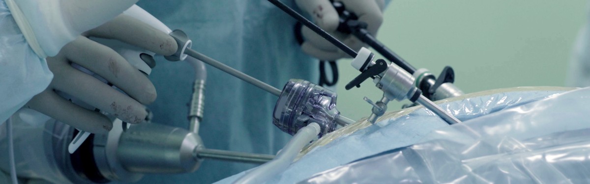
A new project led by Imperial College London could help surgeons to visualise tumours during keyhole surgery, making the treatment more precise.
The researchers, funded by the NIHR Invention for Innovation (i4i) programme, will enhance an existing surgical probe developed by Lightpoint Medical to generate ‘visual heat maps’ of prostate tumours.
By combining robotics and medical imaging, they will develop a system that will highlight cancerous tissue in real-time on screen in the operating theatre.
If successful, in future this minimally invasive tool could help remove cancerous more accurately. This could mean fewer men with prostate cancer need to return to hospital for additional treatment, such as radiotherapy or cancer drugs, which can have a major impact on patients’ quality of life.
Lead researcher Professor Dan Elson, from the Imperial College Institute of Global Health Innovation, said: “To the naked eye, cancerous tissue is virtually impossible to distinguish from healthy tissue, meaning surgeons are often left with difficult and risky decisions on how much to remove.”
“Our research aims to give surgeons the crucial information they need to guide decisions in the operating theatre, which we hope could one day transform the outlook for prostate cancer surgery by reducing side effects and the likelihood that the disease will return.”